The 1MICRON-imaging, an EIC Pathfinder project, develops ultra-high-resolution x-ray sensors for real-time cancer margin detection in surgery, aiming to revolutionize pathology and imaging with faster, more accurate diagnostics.
Discover the project: browse the digital folder or download.
The 1MICRON project aims to revolutionize medical imaging by developing a monolithic x-ray sensor with 1-micron spatial resolution using integrated CMOS electronics and edge-on deep silicon geometry. This high-efficiency sensor enhances phase contrast imaging and allows for real-time, in-surgery cancer margin assessment, potentially eliminating over 100,000 delayed treatments annually in Europe. The technology also opens new markets, including developing countries. A secondary goal is to demonstrate its use in future CT systems, enabling low-power, high-flux imaging and non-invasive “3D virtual biopsies.” The expert European team behind 1MICRON is poised to drive a major shift in pathology and clinical imaging.
Read more about how the new photon-counting CT technology was developed in Stockholm, which forms the basis of this project at the completed research programme: Spectral CT-imaging and Endovascular Techniques.
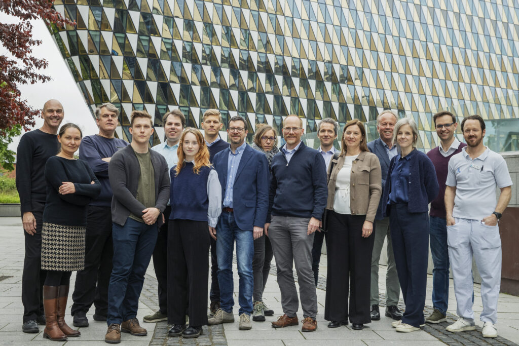
Team
PI´s
Mats Danielsson, KTH
Johan Hartman, Karolinska Institutet
Julia Herzen, TUM
Manuel Rolo, INFN- Instituto Nazionale di Fisica Nucleare
Lucio Pancheri, University of Trento
Anders Björklid, Prismatics Sensors AB
Project Management
Moa Yveborg Tamm, KTH
Public Relations and Communications
Johan Schuber, KTH, jschuber@kth.se, +46 705 510809
Deliverables
The 1MICRON project will deliver a set of key results that document progress and support impact across research, development, and application. These include technical reports, design specifications, experimental validation data, and prototype demonstrators.
Deliverables are aligned with the project’s work packages and track advancement from fundamental development to system integration and real-world validation. In addition, the project will produce dissemination and exploitation outputs to ensure that results are shared and taken forward beyond the project duration.
Public deliverables will be made available to promote transparency and knowledge sharing, while selected outputs may remain confidential to protect intellectual property and support future commercialization.
The project last from 1 March 2024 to 28 February 2029.

The purpose of the programme was to use technology to diagnose and treat cardiovascular diseases, including stroke, and cancer.
The research programme Spectral CT‑imaging and Endovascular Techniques (2019–2023) was led by two internationally recognised Principal Investigators — both also co‑founders of MedTechLabs:
- Professor Mats Danielsson, Professor of Medical Imaging Physics at KTH Royal Institute of Technology – a global pioneer in silicon‑based photon‑counting X‑ray detectors and a driving force behind next‑generation CT technology.
- Professor Staffan Holmin, Professor of Clinical Neuroimaging at Karolinska Institutet and Senior Consultant in Neuroradiology at Karolinska University Hospital – a leading innovator in endovascular techniques, stroke intervention, micro‑biopsy technologies and minimally invasive diagnostics.
Their combined vision and complementary expertise in physics‑driven imaging and advanced clinical interventions formed the foundation of MedTechLabs — and shaped the success of this five‑year programme.
A Completed Programme Defining the Future of CT
The research programme concluded in 2023, marking a major step forward in photon‑counting CT. MedTechLabs’ strategic tenure‑track recruitment strengthened the research environment, including Mats Persson (KTH) whose work on deep‑learning image reconstruction, noise suppression and modelling advanced the capabilities of silicon‑based CT.
Clinical Validation at Karolinska University Hospital
A hallmark achievement was the clinical evaluation of the world’s first silicon‑based photon‑counting CT prototypes, installed at the MedTechLabs CT‑lab in BioClinicum at Karolinska University Hospital. More than a hundred patients across several organ systems have been scanned, generating critical data that supports regulatory approval processes and accelerates technological refinement. This close integration of engineering, clinical expertise and hospital‑based prototyping is a core strength of MedTechLabs.
Breakthroughs in Endovascular Techniques and Stroke Education
Under Staffan Holmin’s leadership, the programme also developed novel endovascular tools, including micro‑biopsy instruments, cell delivery methods, and new approaches to vascular access in the brain and other organs. MedTechLabs further contributed to clinical implementation by developing a widely used education in acute stroke management, focusing on perfusion‑based decision support up to 24 hours after onset. This course now improves stroke diagnostics and treatment pathways across Sweden.
Learn More – Impact Case Available
For those interested in the full research and innovation journey, explore the impact case Better diagnostics with silicon‑based photon‑counting CT, which is available as a browsable online version and also as a downloadable file on this page.
Research Leader Mats Persson, KTH Royal Institute of Technology
In order for the new (photon-counting) computed tomography technology to reach its full potential, the newly developed hardware needs to be supplemented with improved data processing algorithms so that measured data is fully utilized and provides the best possible image quality. In this project, we develop the next generation of image reconstruction methods for photon computing computed tomography and evaluate the resulting image quality. In collaboration with the General Electric Research Center in Niskayuna, NY, USA, we have developed a method for correcting for physical effects when taking pictures that can otherwise incluce artifacts. In collaboration with the Department of Mathematics at KTH, we have also developed an image reconstruction method based on deep neural networks, so-called deep learning, which can greatly reduce the noise in the images. In a few years’ time, the combination of photo-counting computed tomography with the next generation of image reconstruction can take the image quality in computed tomography to a whole new level.
Research leader Mats Persson, KTH
Lung X-rays and computed tomography play a major role in the care of covid-19 patients. At the same time, knowledge on how X-ray images should be interpreted is still limited. The departments of Medical Technology and Medical Image Physics at KTH have in collaboration with Karolinska University Hospital developed a software that largely automates the analysis of X-ray images of covid patients, by marking the lungs and lung damage caused by covid-19. This can save huge amounts of time for physicians who analyze large amounts of patient images to find patterns that can help with treatment. MedTechLabs also develops AI tools based on deep neural networks to automatically analyze computed tomography images of covid patients and predict the patient’s future course of the disease.
Research Leader Niclas Roxhed, KTH Royal Institute of Technology
The project aims to develop extremely small drug capsules, about as thin as a hair, which can be delivered with endovascular techniques. The release of drugs from the capsule is controlled by a remote signal outside the body. This makes it possible to release medication on command, for example once a week or month without the need for a new procedure. To achieve this, we develop capsules based on micro- and nanotechnology that make it possible to construct mechanical structures on a micro-scale and which are activated in a specific way with the help of an external signal. The capsules will be evaluated with a cancer drug and tested in relevant models.
Research Leader Staffan Holmin and Jeroen Goos, Karolinska Institutet
We use immune cells, a poison peptide and bispecific antibodies to specifically target radiation to tumors. Undesirable radioactive radiation is reduced with this targeted method. Protocols for radioactive labeling of immune cells have been optimized and established. The distribution of radiolabeled immune cells (human stromal cells, rat macrophages, human peripheral mononuclear cells) in the body has been monitored in animal models using positron emission tomography (PET) imaging studies. A manuscript with initial results is currently under review. Translation of the results in preparation for clinical trials has begun. In addition, we have radioactively labeled a brain tumor-specific scorpion venom peptide. The radiolabeling was successful and we were able to show that the radiolabeled peptide could bind to glioma cells in the brains of mice. These studies potentially provide new treatment options for cancer patients.
Research Leader Staffan Holmin, Karolinska Institutet
We have continued with the development of the concept of inserting a thin instrument (so-called Extroducer) inside the vessels and have now e.g. succeeded in creating access to the brain parenchyma in pigs via veins. This enables studies on cell transplantation and potentially on sampling. We have published results of heart and kidney access as well as modified technique for access to the pancreas. We have also further developed, tested and patented a microbiopsy tool to enable minimally invasive cardiac sampling for all parts of the heart and developed and verified tissue management protocols for analysis of RNA in these small samples. Ongoing tests of the cardiac biopsy instrument for verification in human tissue are performed in collaboration with Sahlgrenska University Hospital. Development and testing of a microbiops tool for use inside Extroducer is ongoing. We have recently developed and are testing a new instrument dedicated to sampling endothelial cells in various disease states. Furthermore, we have published results of clinical studies of double energy CT scanning after thrombectomy (where blood clots are removed with thin tools via blood vessels) and during thrombolysis (dissolution of blood clots). We have also done a data analysis and published the results from the Stockholm Triage Study, which meant that patients with suspected blood clots in the large vessels of the brain were transported directly to the new Karolinska University Hospital. In addition, we have published new experimental MRI and PET-based concepts to identify endangered brain tissue in acute ischemic stroke.