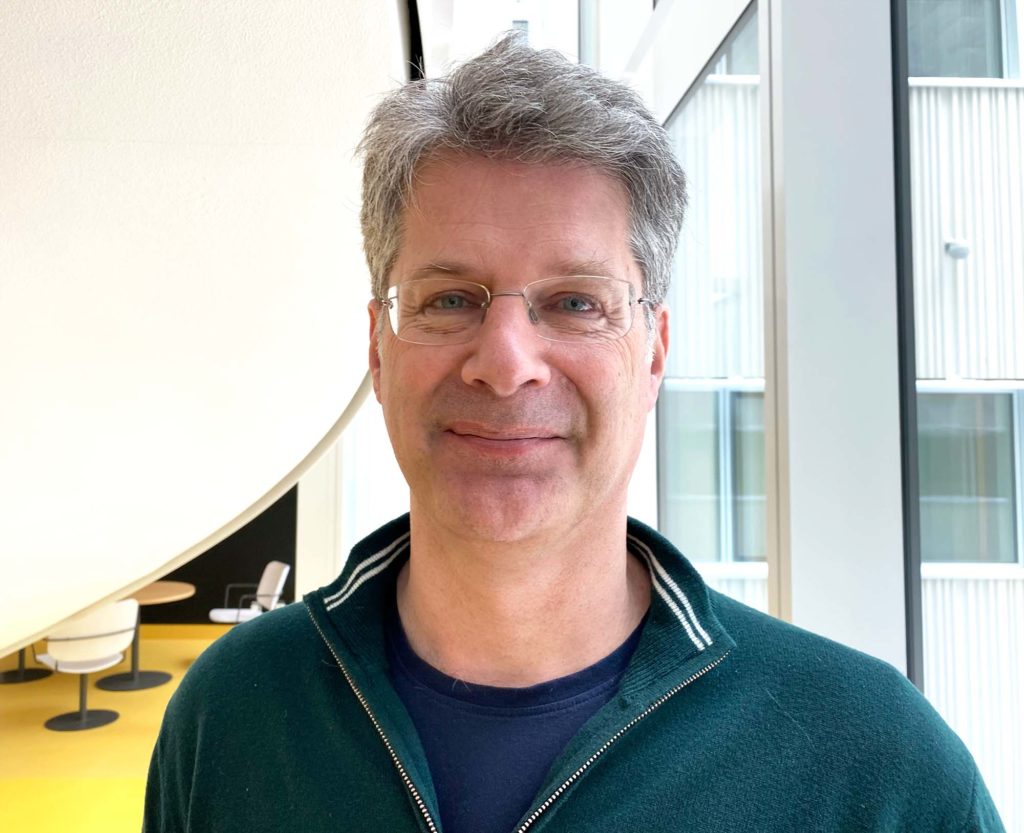
Matilda Larsson is a new member of the MedTechLabs steering group. She works at KTH, where her research focuses on imaging injuries during childbirth. We asked Matilda to introduce herself.
Can you tell us a little about your background and what you do now?
I am a professor of medical imaging at KTH, Department of Medical Engineering and Health Systems, where I am also deputy head and head of the Department of Medical Imaging. I lead a research group in ultrasound imaging, where we develop new methods for measuring the mechanical properties of organs and tissues. Our largest ongoing project focuses on the diagnosis and prevention of birth injuries to the pelvic floor muscles, which is an area where new imaging technology can have great clinical significance.
What did you know about MedTechLabs before you joined the steering group?
Above all, I was aware of several of the exciting and successful projects that have received funding from MedTechLabs over the years. I also perceive MedTechLabs as an important platform for building bridges between academic research, clinical needs, and innovation in medical technology.
What perspectives and ideas do you think you can contribute there?
I hope to contribute a strong research perspective that combines technical development with clear clinical benefits. I am passionate about strengthening the collaboration between technical research and healthcare, and with my background in medical imaging, I want to contribute to the further development of multidisciplinary research and to the translation of new methods into practical solutions that create patient benefits. In addition, I believe that my experience in educating future engineers and researchers in medical technology can be important in creating the conditions and expertise needed for the next generation of innovators to develop and implement new technical solutions in healthcare.
Finally, what role do you think MedTechLabs can and should play in Swedish medtech?
I see MedTechLabs as a key player in driving Swedish medical technology research forward and as a continued strong engine for future breakthroughs in medical technology in Sweden. Through its model for how academia and healthcare collaborate to create world-class research on clinically relevant problems, the center can help meet some of healthcare’s biggest challenges and ensure that Sweden remains at the forefront of medical technology innovation.
– Interdisciplinary and industrial experience gives Moa Yveborg Tamm confidence as the EU project 1Micron gets underway.
1Micron is the flagship project funded by the EU’s EIC Pathfinder Open programme to develop an ultra-high resolution X-ray sensor for real-time detection of cancer margins in surgical procedures. If successful, the project could revolutionise current pathology and imaging. Moa Yveborg Tamm is coordinating the research, which is being carried out by researchers in three countries. Moa is returning to academia after several years working in industry at GE Healthcare.
Hi Moa, what are you up to right now?
“We have just started creating the project, which is based on the participation of several partners. Now it’s about getting everyone into 1Micron and we have a kick-off already on 22 May. Then everyone will meet together for the first time, and we can start discussing the technical aspects together. At this stage, you don’t know how much control is needed, it will only become clearer when the discussion on deliverables, decisions and forms of cooperation is actively pursued.”
How many people are actually involved in 1Micron?
“In addition to Mats Danielsson and Mats Persson from KTH Royal Institute of Technology and Johan Hartman from KI, the Italian nuclear physics institute INFN, the University of Trento and the Technical University of Munich (TUM) are involved. In addition, there are industrial partners such as Prismatics Sensors AB. Our partners are a key to the success we hope for. For example, the medical researchers are helping with validation so that we can understand how and in what way the new sensor we will develop can make a difference in clinical applications.”
The interdisciplinary nature of the project and its industrial links make it complex?
“Yes, and here I feel it is good that I have learnt what it is like to work interdisciplinary and in industry. In my experience, it is often necessary to have a systems approach right from the start. It sets some of the framework for how you need to rig the project and the importance of not locking yourself into a solution that is not scalable early on. It’s okay not to have all the answers from the start, as long as you have really good people involved in the project. Which we do.”
It still sounds like a tough assignment, why did you take it?
“Why did you take it? ‘It’s a super fun project, something that could revolutionise medical imaging. A bit like what we’ve done before with the development of the first photon-counting CT with silicon detectors, but still completely new. There is the potential to do something amazing and with very exciting technology. Plus, I get to work with really good partners who know their fields. I would say that we put together the perfect team with 1Micron!”
And what happens after the kick-off on 22 May?
“We have already started working towards the deliverables we have set up, but now the work is intensifying. A key piece is the sensor design and showing how it can achieve the phase contrast we are aiming for, in real time. One of the early specifications will revolve around this, along with the other hardware needed to enable phase contrast. Another thing that will happen this year is that Karolinska Institutet and TUM will provide us with a baseline to compare against, by collating how well current digital detectors perform.”
What are you most looking forward to in the project?
“I think I’m looking forward to being part of the biggest thing to happen in medical imaging in a long time. It will of course be a challenge, but we have physics on our side and a hand-picked team, which also gives us really good odds. What I also appreciate is that we have a partner from industry who knows a lot about the technical requirements of what will then be out in the hospitals. An ‘insider’, you might say. You don’t always have that in academic research projects, and it’s also something that suggests that 1Micron can achieve its goals.”
Read more about the project on the 1MICRON website
A new centre that brings together current and future expertise and technology in medical imaging. Expectations for the Centre of Imaging Research (CIR) and what it can achieve in terms of research, industry collaboration and clinical application are sky-high. In this article, Daniel Lundqvist, who heads the centre, talks about how the CIR will make its mark in Sweden and internationally.
Daniel Lundqvist formally became director of CIR - Centre for Image Research - in October 2022. The importance of the new centre was marked by the participation of both Karolinska Institutet's President Ole Petter Ottesen and Karolinska University Hospital's CEO Björn Zoëga in the inauguration. Most of the centre is in Karolinska University Hospital's BioClinicum research building, which is also well integrated with KI. MedTechLab's CT lab is now part of the CIR infrastructure. One block away is the Scilifelab, which also conducts research in the field of imaging.

Daniel Lundqvist is a brain researcher at KI and has a passion for medical infrastructure. He explains that the first thoughts of a single imaging centre came more than 20 years ago, and that brain researcher Martin Ingvar, former head of the MR Centre, and Staffan Holmin, physician and research leader at MedTechLabs, were among those who started working on realising the ideas early on.

The CIR will bring together the best instruments, platforms, and technologies available in medical imaging, and Daniel has started building the centre's structure and initial operations.

– In addition to the co-location, we have upgraded with the latest in medical imaging technology. But I want to emphasise that as important as it is to have all this great technology in one place, it is equally important to bring together all the experts in the field of imaging so that they can collaborate and learn from each other, Daniel explains.

By bringing these people together, level four of the BioClinicum also forms a professional community that is unparalleled in the world. The CIR is likely to be attractive to researchers internationally, which will benefit recruitment to research projects linked to the centre.

One goal of the CIR is to create benefits for users in research, healthcare and industry. The centre makes it possible to devote resources to their common needs, such as a new platform for storing and sharing image data, which would have been difficult for the various users to create on their own.

The activities at CIR will contribute to the translation of research results into clinical activities, known as translational research. This is facilitated by the proximity to Karolinska University Hospital, which is just a stone's throw away with its doctors and patients.

CIR is partly based on imaging facilities that have already existed for a long time at Karolinska Institutet and Solna University Hospital. What is new is that these are being brought together physically. An important reason for establishing the centre was the realisation of how important it is to be able to perform multimodal imaging, i.e., to combine different types of imaging. Today, this is a logistical challenge when facilities are scattered in different locations, and it also puts a strain on patients or research subjects when they must be moved around. Surprisingly rarely, for example, MRI, MEG, PET and CT are combined on the same individual, even though it is often important to obtain a detailed and multifaceted overall picture. The CIR will therefore enable a wide range of imaging of the structure, function, and metabolism of living organs, using the various modalities of technology available and constantly evolving. The diagnosis and study of cancer, cardiovascular diseases and brain imaging are the focus. For example, NatMEG, a national infrastructure for magnetic encephalography that is one of the most advanced in the world, will be part of the centre.

One of the CIR's strengths is that we have the very latest or even tomorrow's methods that we are evaluating and trying to utilise. Such as the new photon counting CT technology with silicon detectors that is now being validated in MedTechLabs and a new MRI with ultra-high field strength, called 7-tesla, which is available in Huddinge. - We are currently upgrading NatMEG with an improved system for measuring brain activity with a 128-channel system for on-scalp MEG. This is the latest and greatest in the world," says Daniel.

The centre's ability to provide researchers and clinicians with more information about the appearance, function and metabolism of organs can be of great importance for individual patients with severe diseases. Daniel takes epilepsy as an example:

- In the brain, epileptic seizure activity can begin as an intense activation of a very small cortical region, which then expands over time to involve progressively larger and larger cortical areas. If a patient needs surgery to remove the part of the brain where seizure activity starts, one will naturally want to identify the exact starting point as accurately as possible. With the technology available at CIR and NatMEG, we believe that it will be possible to find the seizure activity’s starting point more accurately and thus the method can also guide the neurosurgeon with greater certainty, says Daniel.

Another exciting area where CIR could play an important role is theranostics, which combines therapy and diagnostics. Theranostics uses a targeted radiopharmaceutical that can identify and diagnose the extent of a cancer, and then treat the cancer either with a therapeutic radiopharmaceutical (or with targeted oncological treatment).  This method successfully improves the effectiveness of cancer treatment while maintaining quality of life. The field is relatively new and rapidly developing. Sweden has an internationally prominent position in the field, and is among the most active countries in Europe, both in the preclinical development of theranostic drugs to translating and participating in clinical theranostic studies and in initiating them.

Nationally, there are many links to CIR and Daniel is already involved in several Swedish coordination projects where the focus is on sharing and making data available. Internationally, the centre will also be an important Swedish node for various research collaborations, such as the European e-infrastructure for brain research, EBRAINS. Synchronising CIR with EBRAINS will make it easier for CIR's imaging platforms and users to share and use data in European collaborations between researchers, clinicians, and industry.

- It has taken a while to realise what a great job I have been given and I am both proud and humbled by this assignment. Since I have worked with imaging issues and imaging infrastructure for a long time, I know that it can be challenging and involves a lot of responsibility. But it is so exciting to be involved in creating something that offers new and unique opportunities that will ultimately benefit healthcare and patients. I share this feeling with many others who will work here at CIR, concludes Daniel.