Research leader Mats Persson, KTH
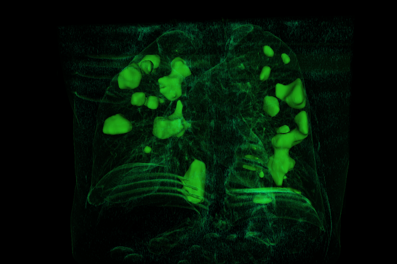
Lung X-rays and computed tomography play a major role in the care of covid-19 patients. At the same time, knowledge on how X-ray images should be interpreted is still limited. The departments of Medical Technology and Medical Image Physics at KTH have in collaboration with Karolinska University Hospital developed a software that largely automates the analysis of X-ray images of covid patients, by marking the lungs and lung damage caused by covid-19. This can save huge amounts of time for physicians who analyze large amounts of patient images to find patterns that can help with treatment. MedTechLabs also develops AI tools based on deep neural networks to automatically analyze computed tomography images of covid patients and predict the patient’s future course of the disease.
We use cookies to improve your user experience and to collect information about your visit. You can read more about our cookies and change your settings here. Read more
Cookies save information about how you use the website, data that can be reused. Read more
Necessary for the website to function.
Read moreThese cookies are necessary for our website to function and therefore cannot be turned off. They are used, for example, when you set personal preferences, log in or fill out a form. You can set your browser to block or warn you about these cookies, but some parts of the website will not work then.
close-cookie-bar
wants-ec-cookies
wants-fc-cookies
wants-mc-cookies
wants-ac-cookies
For some features on our website.
Read moreThese cookies make it possible to provide improved functionality and customization on our website. If you do not allow these cookies, some functions may not work correctly.
YSC
Measures user patterns and creates statistics.
Read moreThese cookies allow us to count the number of visits and traffic sources so that we can measure and improve our website. The information these cookies collect is completely anonymous. If you do not allow these cookies, we will not know when you have visited our website.
_ga_S8QD52ZQ5E
_ga
_gid
Sign up for MedTechLabs newsletter, and you won’t miss any news or events from us. The newsletter is in english and distributed on a regular basis.